HOME >> 海外出版社刊 洋書販売 新着書籍 >>
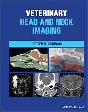
Veterinary Head and Neck Imaging

Wiley-Blackwell
| Author | : | Peter V. Scrivani |
価格:34,210円 (本体 31,100円+税) 送料サービス
・Release: 2022
・ISBN: 9781119118596
・800 Pages
・Trim Size: 226.1 X 35.6 X 281.9 ・Hardcover
Description
A complete, all-in-one resource for head and neck imaging in dogs, cats, and horses
Veterinary Head and Neck Imaging is a comprehensive reference for the diagnostic imaging of the head and neck in dogs, cats, and horses. The book provides a multimodality, comparative approach to neuromusculoskeletal, splanchnic, and sense organ imaging. It thoroughly covers the underlying morphology of the head and neck and offers an integrated approach to understanding image interpretation.
Each chapter covers a different area and discusses developmental anatomy, gross anatomy, and imaging anatomy, as well as the physical limitations of different modalities and functional imaging. Commonly encountered diseases are covered at length.
Veterinary Head and Neck Imaging includes all relevant information from each modality and discusses multi-modality approaches. The book also includes:
- A thorough introduction to the principles of veterinary head and neck imaging, including imaging technology, interpretation principles, and the anatomic organization of the head and neck
- Comprehensive explorations of musculoskeletal system and intervertebral disk imaging, including discussions of degenerative diseases, inflammation, and diskospondylitis
- Practical discussions of brain, spinal cord, and cerebrospinal fluid and meninges imaging, including discussions of trauma, vascular, and neoplastic diseases
- In-depth treatments of peripheral nerve, arterial, venous and lymphatic, respiratory, and digestive system imaging
Veterinary Head and Neck Imaging is a must-have resource for veterinary imaging specialists and veterinary neurologists, as well as for general veterinary practitioners with a particular interest in head and neck imaging.
Table of contents
Preface
SECTION 1 INTRODUCTION TO HEAD AND NECK IMAGING IN ANIMALS
1 Some Basic Concepts about Head and Neck Anatomy
1.1 Terms of Location, Orientation, and Movement
1.2 External Features of the Head and Neck
1.3 Overview of Neuroanatomic Localization during Neuroimaging
1.3.1 Divisions of the Central Nervous System
1.3.2 Neuroaxis Localization
1.3.3 Clinical Descriptors for the Location of Intracranial Abnormalities
References
2 Some Basic Concepts about Medical Imaging
2.1 Introduction
2.1.1 What is an Image?
2.1.2 What is Medical Imaging?
2.2 Medical Imaging Devices
2.2.1 Imaging Technologies
2.2.2 Imaging Techniques, Applications, and Examinations
2.3 The Medical Image
2.3.1 Picture Elements and Volumetric Picture Elements
2.3.2 Representing Tissue Characteristics through the Grayscale
2.3.3 Resolution
2.4 Image Evaluation
2.4.1 Getting Started
2.4.2 Imaging Signs and Patterns
2.4.3 Image Evaluation
References
SECTION 2 MUSCULOSKELETAL IMAGING
3 The Musculoskeletal System
3.1 Imaging Anatomy
3.1.1 Bone
3.1.2 Joints and Ligaments
3.1.3 Muscles and Tendons
3.1.3.1 Fascia and Fascial Compartments
3.2 Musculoskeletal Abnormalities
3.2.1 Developmental Malformations
3.2.1.1 Cranium, Face, and Craniocervical Junction
3.2.1.2 Vertebrae
3.2.2. Degenerative Diseases
3.2.2.1 Joints
3.2.2.2 Vertebrae
3.2.3 Inflammatory Diseases
3.2.3.1 Infectious
3.2.3.2 Non-infectious
3.2.4 Neoplasia
3.2.5 Nutritional, Metabolic, Toxic Diseases
3.2.6 Trauma
3.2.6.1 Soft-tissue Trauma
3.2.6.2 Fracture
3.2.6.3 Dislocation
References
4 Intervertebral Disks
4.1 Imaging Anatomy
4.2 Intervertebral Disk Abnormalities
4.2.1 Developmental Malformations
4.2.2 Infection/Inflammation
4.2.3 Trauma
4.2.4 Degeneration
4.2.5 Herniation
References
SECTION 3 NERVOUS SYSTEM IMAGING
5 Cerebrospinal Fluid
5.1 Imaging Anatomy
5.2 CSF Production, Absorption, and Flow
5. 3 Cerebrospinal Fluid Abnormalities
5.3.1 Intra-axial Fluid Accumulations
5.3.2 Extra-axial Fluid Accumulations
5.3.3 Intramedullary Fluid Accumulations
5.2.4 Extramedullary Fluid Accumulations
References
6 The Central Nervous System
6.1 Imaging Anatomy
6.2 Brain and Spinal Cord Abnormalities
6.2.1 Imaging Patterns of CNS Disease
6.2.1.1 Some Additional Imaging Signs
6.2.1.2 Contrast Enhancement
6.2.2. Secondary Intracranial Abnormalities
6.2.2.1 Intracranial Hypertension
6.2.2.2 Cerebral Edema
6.2.2.3 MRI Signs Induced by Seizures
6.2.2.4 Brain Herniation
6.2.2 Developmental Malformations
6.2.3 Vascular Disorders
6.2.3.1 Ischemia
6.2.3.2 Hemorrhage
6.2.3.3 Hemorrhagic Infarction
6.2.4 Trauma
6.2.4.1 Traumatic Brain Injury
6.2.4.2 Traumatic Spinal Cord Injury
6.2.5 Neoplasia
6.2.6 Inflammatory Diseases
6.2.6.1 Infectious
6.2.6.2 Non-Infectious
6.2.7 Degenerative Diseases
References
7 The Peripheral Nervous System
7.1 Imaging Anatomy
7.1.1 Cranial Nerves
7.1.2 Spinal Nerves
7.1.2.1 The Cervical Nerves
7.1.2.2 The Brachial Plexus
7.1.2.3 The Sympathetic Division
7.2 Peripheral Nerve Abnormalities
7.2.1 Neoplasia
7.2.2 Trauma
7.2.3 Inflammatory Diseases
7.2.4 Compression
7.2.1 Degenerative Diseases
References
SECTION 4 SPLANCHNIC (VISCERA), VASCULAR, AND SENSE ORGAN IMAGING
8 The Digestive System
8.1 Imaging Anatomy
8.1.1 Oral Cavity
8.1.2 Pharynx
8.1.3 Cervical Esophagus
8.2 Digestive Track Abnormalities
8.2.1 Developmental Malformations
8.2.2 Dysphagia
8.2.3 Neoplasia
8.2.4 Inflammation
References
9 The Respiratory System
9.1 Imaging Anatomy
9.1.1 Nasal Cavities and External Nose
9.1.2 Paranasal Sinuses
9.1.3 Nasopharynx, Larynx, and Cervical Trachea
9.2 Respiratory Track Abnormalities
9.2.1. Developmental Anomalies
9.2.2. Inflammation/Infection
9.2.3. Neoplasms
9.2.4. Degenerative Disorders
References
10 Sense Organs, Circulatory System, and Endocrine System
10.1 Imaging Anatomy
10.1.1 Eye
10.1.2 Ear
10.1.3 Circulatory System
10.1.4 Endocrine System
10.2 Orbital Disorders
10.2.1 Trauma
10.2.2 Inflammatory Disease
10.2.3 Neoplasms
10.3 Ear Disorders
10.3.1 Ear Diseases
10.3.2 Guttural Pouch Disease
10.3.3 Imaging Patterns of Disease
10.4 Circulatory and Endocrine Disorders
10.4.1 Developmental Anomalies
10.4.2 Endocrine Disorders
10.4.3 Circulatory System Disorders
References
Index


