HOME >> 海外出版社刊 洋書販売 新着書籍 >>
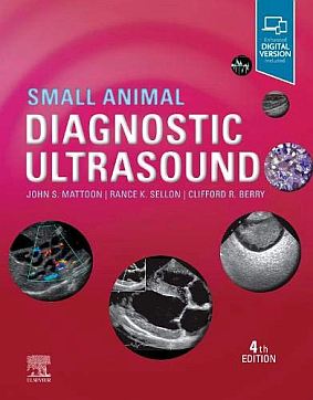
Small Animal Diagnostic Ultrasound, 4E

Elsevier
| Author | : | John S. Mattoon / Rance K. Sellon / Clifford Rudd Berry |
価格:29,040円 (本体 26,400円+税) 送料サービス
・Release: 2020
・ISBN: 9780323533379
・752 Pages
・Trim Size: 224 X 32 X 284 ・Approx. 800 illustrations (800 in full color)
Description
Every clinician that has an interest in veterinary diagnostic imaging should have this reference! Small Animal Diagnostic Ultrasound 4th Edition provides in-depth coverage of the latest techniques applications and developments in veterinary ultrasonography. It shows how ultrasonography can be an indispensable part of your diagnostic workup for everything from cardiac and hepatic disease to detached retinas and intestinal masses. All-new content on internal medicine is integrated throughout the text addressing disease processes and pathologies their evaluation and treatment. Written by expert educators John S. Mattoon Rance K. Sellon and Clifford R. Berry this reference includes access to an Expert Consult website with more than 100 video clips and a fully searchable version of the entire text.
New to this edition
- NEW! Updated content on diagnostic ultrasound ensures that you are informed about the latest developments and prepared to meet the challenges of the clinical environment.
- NEW! Coverage of internal medicine includes basic knowledge about a disease process, the value of various blood tests in evaluating the disease, as well as treatment strategies.
- NEW editors Rance K. Sellon and Clifford R. Berry bring a fresh focus and perspective to this classic text.
- NEW! Expert Consult website includes a fully searchable eBook version of the text along with video clips demonstrating normal and abnormal conditions as they appear in ultrasound scans.
- NEW! New and updated figures throughout the book demonstrate current, high-quality images from state-of-the-art equipment.
- NEW contributing authors add new chapters, ensuring that this book contains current, authoritative information on the latest ultrasound techniques.
Key Features
- Logical organization makes reference quick and easy, with chapters organized by body system and arranged in a head-to-tail order.
- Coverage of Doppler imaging principles and applications includes non-cardiac organs and abdominal vasculature.
- Photographs of gross anatomic and pathological specimens accompany ultrasound images, showing the tissues under study and facilitating a complete interpretation of ultrasound images.
- More than 100 video clips demonstrate normal and abnormal conditions as they appear in ultrasound scans, including conditions ranging from esophageal abscess to splenic hyperplasia.
- More than 2,000 full-color images include the most current ultrasound technology.


